Reservoirs can be filled with fluid or mock blood to represent pleural effusion
Bilateral chest drain and needle decompression pads
REALISM
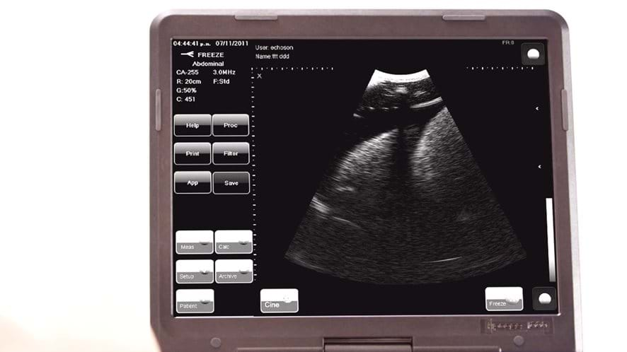
Can give the impression of breathing under ultrasound when using the advanced pad
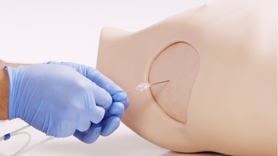
Needle decompression air reservoirs provide realistic release of air on insertion of needle
VERSATILITY
Suitable for supine, sitting or leaning forwards positions
Works with thoracic seals when using the standard pad
Affordable replaceable pads
SAFETY
Latex free
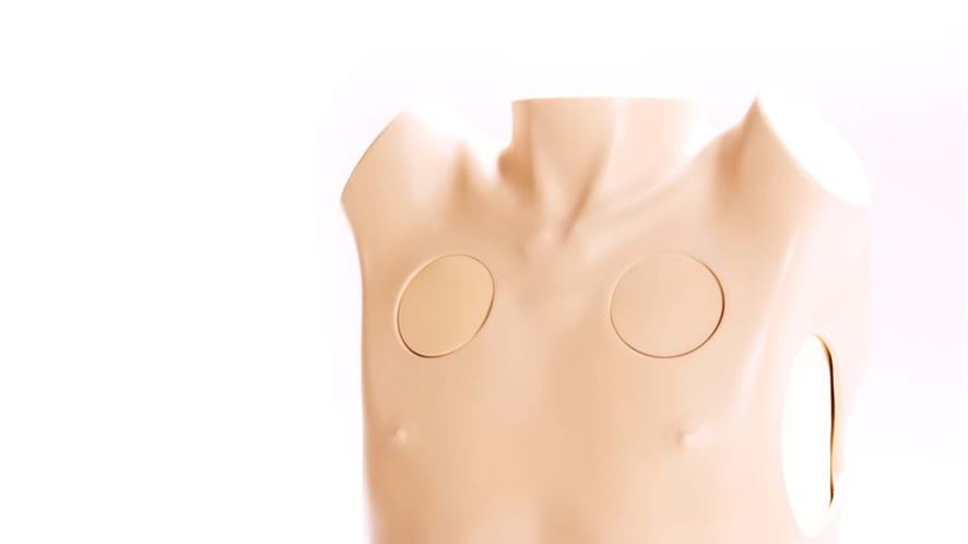
ANATOMY
Representation of adult male thorax with arms raised
Bony and soft tissue landmarks: manubriosternal joint, clavicles, ribs, pectoralis major and latissimus dorsi
Internal ultrasound anatomy: diaphragmatic structures and collapsed lung
SKILLS GAINED
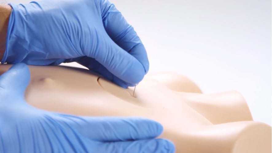
Needle decompression of a tension pneumothorax (at both the 2nd and 5th intercostal space)
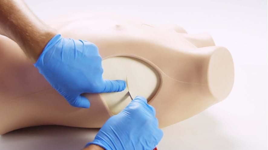
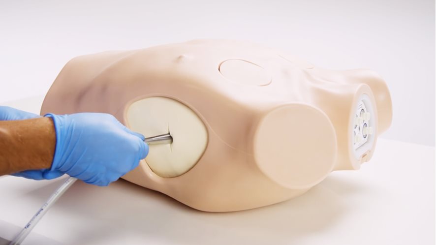
Open, or cut-down chest drain insertion: recognition of correct position, surgical incision, blunt dissection through chest wall, perforation of pleura and finger sweep
Suture of tube to chest wall
Ultrasound-guided chest drain insertion (Seldinger-type), including insertion of needle under direct vision and ultrasonic recognition of chest structures